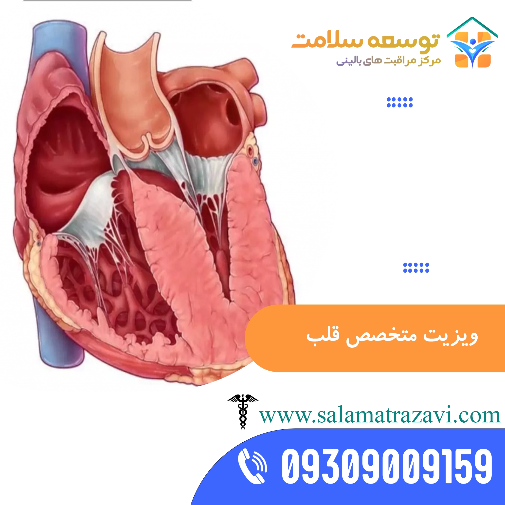
Description
Cardiologist at home in Mashhad
Heart failure is a progressive, systemic disorder that affects not only the heart’s ventricles but also the kidneys, lungs, liver and peripheral circulation. A clear understanding of how it develops is fundamental for the cardiologist managing a patient at home.
When the heart muscle (myocardium) is damaged by a heart attack, long-standing high blood pressure or cardiomyopathy, cardiac output falls. This fall in stroke volume immediately activates the body’s compensatory mechanisms. In the short term these help maintain blood flow to vital organs such as the brain and the heart itself, but in the long term they have harmful effects.
The sympathetic nervous system is one of the first to be activated. Release of noradrenaline and adrenaline increases heart rate and contractility and narrows the peripheral blood vessels to maintain blood pressure. However, a constantly raised heart rate increases the heart muscle’s oxygen demand and shortens the time for the ventricles to fill in diastole, which worsens ischaemia in the ventricular walls.
At the same time, the renin–angiotensin–aldosterone system (RAAS) is activated. Reduced blood flow to the kidneys and reduced sodium delivery to the macula densa cause renin to be released from the juxtaglomerular cells. Renin converts angiotensinogen to angiotensin I, which angiotensin-converting enzyme (ACE) converts to angiotensin II.
Angiotensin II is a very powerful vasoconstrictor that greatly increases systemic vascular resistance (afterload).
It also stimulates the adrenal cortex to release aldosterone, causing the kidneys to retain sodium and water and increasing circulating blood volume (preload). Higher preload and afterload put the ventricular walls under severe strain.
This constant wall stress triggers ventricular remodelling: heart muscle cells (cardiomyocytes) undergo pathological hypertrophy and fibrous tissue increases. These structural changes reduce the ventricle’s elasticity, leading to diastolic dysfunction (inability to relax and fill) and eventually systolic dysfunction.
At home, the cardiologist breaks this harmful cycle by carefully assessing blood biomarkers such as NT-proBNP, which is released in response to stretching of the ventricular wall, and adjusting doses of ACE inhibitors, angiotensin receptor blockers (ARBs) and aldosterone antagonists (such as spironolactone).
Portable and tissue Doppler echocardiography for diagnosing diastolic and systolic heart failure at the bedside
Portable echocardiography at home is more than simple imaging; it acts as an advanced diagnostic laboratory at the bedside.
In heart failure with preserved ejection fraction (HFpEF), also known as diastolic heart failure, the left ventricular ejection fraction (LVEF) may be normal or near normal (above 50%), yet the patient has severe breathlessness and pulmonary oedema. Here the cardiologist uses advanced Doppler techniques to examine how the left ventricle fills.
Blood flow velocity across the mitral valve is measured in early diastole (the E wave) and during atrial contraction (the A wave). Normally E is greater than A, but when relaxation is impaired the ratio reverses (E/A < 1).
In more advanced disease, a pseudonormal or restrictive filling pattern develops, indicating markedly raised left ventricular filling pressures. Using tissue Doppler imaging (TDI), the doctor measures the velocity of the mitral annulus (the e’ wave). The E/e’ ratio is a very useful estimate of pulmonary capillary wedge pressure (PCWP); values above 14 indicate markedly raised filling pressure and a high likelihood of acute lung congestion.
In heart failure with reduced ejection fraction (HFrEF), portable echocardiography helps measure the ejection fraction accurately, assess dyssynchrony of wall contraction and identify areas of abnormal wall motion (hypokinesia, akinesia, dyskinesia) from previous heart attacks. Examining the heart valves with colour Doppler to detect secondary valve leaks (such as functional mitral regurgitation caused by left ventricular dilatation) is another vital step.
This detailed information allows the doctor to decide on the need for advanced treatments such as cardiac resynchronisation therapy (CRT) or implantable cardioverter-defibrillators (ICD) without having to move a weak, very ill patient to hospital.
Advanced 12-lead ECG interpretation at home and distinguishing arrhythmias
Recording and interpreting an electrocardiogram (ECG) at home is one of the cardiologist’s key tools for rapidly assessing the heart’s electrical activity. During the visit, the doctor looks not only for signs of acute ischaemia but also for complex ventricular and supraventricular arrhythmias that can destabilise the circulation. Assessing heart rhythms requires careful analysis of intervals and wave shapes.
In distinguishing wide-complex tachycardias (QRS > 120 ms), the doctor must differentiate ventricular tachycardia (VT) from supraventricular tachycardia with aberrant conduction (SVT with aberrancy). The Brugada criteria are used at the bedside:
– Absence of an RS complex in all precordial leads (V1–V6): if absent, the diagnosis is VT.
– If an RS complex is present, measure the interval from the start of the R wave to the deepest point of the S wave (RS interval); if it exceeds 100 ms in any lead, VT is indicated.
– Look for atrioventricular (AV) dissociation, in which P waves move independently of the QRS complexes; this is the strongest sign of VT.
– Assess the QRS morphology in leads V1 and V6.
In atrial fibrillation, it is important to look for a fibrillating baseline with no P waves and completely irregular R–R intervals. By interpreting these findings and checking the QT interval (especially in patients taking antiarrhythmic drugs such as amiodarone or sotalol), the cardiologist at home helps prevent dangerous arrhythmias such as torsades de pointes and adjusts doses safely at home.
Managing complex medicine regimens and drug interactions in older heart failure patients
Treatment of heart failure involves several classes of medicine at once, each targeting a different mechanism. This polypharmacy carries a high risk of interactions and side effects, especially in older patients who also have chronic kidney disease, diabetes or arthritis. The cardiologist at home monitors and personalises this regimen carefully.
First-line medicines include angiotensin receptor–neprilysin inhibitors (ARNIs) such as sacubitril/valsartan, which now often replace ACE inhibitors. By blocking neprilysin, they raise natriuretic peptide levels and strengthen the body’s natural blood-vessel-widening and diuretic effects.
Starting this medicine requires careful monitoring of blood pressure and potassium. Beta-blockers (such as carvedilol, metoprolol succinate and bisoprolol) are used to control heart rate and reduce the toxic effects of catecholamines. They must be started at very low doses because early on they may reduce contractility and temporarily worsen symptoms.
Sodium–glucose co-transporter 2 (SGLT2) inhibitors such as empagliflozin and dapagliflozin have transformed heart failure treatment. By removing glucose and sodium in the urine, they reduce preload and afterload and protect the heart and kidneys.
In older people, monitoring kidney function (eGFR) and electrolytes (especially potassium and sodium) while taking loop diuretics (such as furosemide) is vital. A common interaction is taking NSAIDs for joint pain, which can cancel the effect of diuretics and, through salt and water retention and narrowing of the kidney’s blood vessels, cause acute kidney injury and worsening heart failure. The cardiologist at home identifies these interactions and prescribes safe alternatives.
Monitoring laboratory biomarkers at home and interpreting trends
Assessing a heart patient’s biochemistry through blood tests at home is a foundation of treatment. The doctor looks not only at single values but at trends over time. Key biomarkers include:
– B-type natriuretic peptide (BNP) and NT-proBNP: released in response to increased pressure and stretch in the ventricles. A sudden rise indicates fluid build-up before obvious symptoms such as severe breathlessness appear, alerting the doctor to increase the diuretic dose.
– Creatinine and blood urea nitrogen (BUN): in heart failure, reduced cardiac output lowers blood flow to the kidneys (cardiorenal syndrome). Rising creatinine and BUN-to-creatinine ratio indicate reduced filtration and kidney venous congestion, requiring medicine doses to be adjusted.
– Electrolytes (sodium, potassium, magnesium): loop diuretics cause loss of potassium and magnesium, while ACE inhibitors and spironolactone retain potassium. Both low potassium (hypokalaemia) and high potassium (hyperkalaemia) can cause dangerous ventricular arrhythmias, so careful, regular monitoring at home is essential for safety.
– Cardiac troponins (cTnI/cTnT): when acute coronary syndrome is suspected, high-sensitivity troponin (hs-cTn) testing helps detect ischaemic heart muscle damage quickly.
Cardiac physiology and occupational therapy in activity tolerance training
Rehabilitation and occupational therapy exercises for heart patients at home must be designed with a full understanding of the circulation’s limits. When an occupational therapist practises daily activities (ADLs) with the patient, specific changes occur in heart parameters that need supervision by the cardiologist.
Activities are classified by metabolic equivalents (METs). One MET equals an oxygen consumption of 3.5 ml per kilogram of body weight per minute at rest. Basic activities such as dressing or brushing teeth need 1.5–2.5 METs, while showering or climbing stairs can raise the requirement to 4–5 METs. In a damaged heart, a sudden rise in METs can make the heart muscle’s oxygen demand exceed supply, causing signs of ischaemia (such as angina or ST depression on the ECG).
During activity training, blood pressure and heart rate responses must be monitored closely. Normally, as exercise intensity increases, systolic blood pressure rises steadily while diastolic pressure changes little or falls slightly. Abnormal responses include:
– a fall in systolic blood pressure (more than 10 mmHg below baseline as activity increases), showing that the left ventricle cannot maintain output under physical stress; a very serious warning sign;
– a marked rise in diastolic blood pressure (above 110 mmHg), indicating severe vasoconstriction and increased afterload;
– failure of the heart rate to rise in line with activity (chronotropic incompetence), which may be caused by high doses of beta-blockers or sinus node disease.
By setting a safe target heart rate through clinical assessment, the cardiologist allows the occupational therapist to design exercise programmes within safe limits.
Energy conservation strategies and adapting daily activities for heart patients at home
A key part of cardiac rehabilitation at home is teaching energy-saving methods that reduce the heart’s workload during daily tasks, so patients can stay independent despite reduced heart function:
– Organising the environment: placing frequently used items in the kitchen and bedroom between shoulder and hip height, to minimise bending or reaching overhead (which increases the heart’s work through contraction of the large shoulder muscles).
– Suitable postures: doing tasks sitting rather than standing; sitting reduces energy use by up to 25%. For example, a shower chair prevents the drop in blood pressure caused by skin blood vessels widening in hot water and prolonged standing.
– Breathing control: teaching pursed-lip breathing during heavier tasks such as standing up from a chair or climbing stairs, which keeps the airways open and reduces the work of breathing.
– Pacing: dividing large tasks into smaller parts with planned rest breaks so the heart rate can return to baseline.
Advanced care at home after coronary bypass surgery (CABG) and valve replacement
Recovery after major heart surgery such as coronary artery bypass grafting (CABG) or valve replacement is a critical period, and managing it at home requires great expertise. After discharge, patients face many physiological and structural challenges. One of the most important is care of the sternotomy (the cut through the breastbone), which needs 6 to 8 weeks to heal fully. During this time the cardiologist at home monitors:
– Stability of the breastbone: examining for abnormal movement or clicking at the sternum, which suggests failure to heal (sternal instability).
– Prevention of wound infection (mediastinitis): checking for local inflammation, pus, fever and increasing pain; deep bone infection needs urgent surgery.
– Post-operative arrhythmias: atrial fibrillation (POAF) occurs in about 30% of patients after CABG. The doctor manages it by adjusting medicines (such as beta-blockers or amiodarone) and correcting electrolytes.
– Anticoagulation: in patients with a mechanical valve, warfarin and close INR (international normalised ratio) monitoring weekly or every two weeks at home help prevent clots on the valve or internal bleeding.
Combined lung and breathing rehabilitation in patients with heart failure and COPD
Many heart patients also have chronic lung disease such as COPD (cardiopulmonary overlap). In these patients it can be hard to tell whether breathlessness comes from heart congestion or bronchospasm. The cardiologist at home distinguishes and treats both by:
– Careful listening to the lungs: distinguishing crackles (fluid in the air sacs from heart failure) from wheezes (narrowed airways in COPD).
– Pulse oximetry at rest and during activity: a sudden drop in oxygen during activity may indicate a need for supplemental oxygen at home.
– Incentive spirometry and deep-breathing exercises: to prevent atelectasis (collapse of parts of the lung), especially after heart surgery.
– Careful medicine adjustment: avoiding non-selective beta-blockers (such as propranolol), which can cause severe bronchospasm in COPD, and using highly cardioselective beta-blockers such as bisoprolol or nebivolol instead.
Assessing peripheral arterial disease (PAD) and venous disease in heart patients
Arterial and venous diseases of the legs often accompany heart disease and can cause similar symptoms. At a home visit, the cardiologist assesses:
– Peripheral arterial disease (PAD): narrowing of the leg arteries by atherosclerosis, causing pain on walking (intermittent claudication). The doctor feels the foot pulses (dorsalis pedis and posterior tibial) and measures the ankle–brachial index (ABI) at home with a portable Doppler probe to assess severity.
– Deep vein thrombosis (DVT): a clot in the deep veins of the leg, with a risk of travelling to the lungs and causing pulmonary embolism (PE). The leg is examined for one-sided swelling, warmth, redness and pain.
– Chronic venous insufficiency (CVI): damage to the vein valves causing blood to pool in the legs, chronic swelling, skin colour changes (haemosiderin pigmentation) and venous ulcers. Distinguishing this swelling from heart failure oedema is vital for correct treatment (compression stockings rather than diuretics).
Acute coronary syndromes (ACS): first supportive steps at home
Acute coronary syndromes (such as unstable angina and acute myocardial infarction) need immediate transfer to a centre with emergency angioplasty (primary PCI). If you suspect a heart attack, call the emergency services (115) immediately. Direct involvement of a cardiologist in the first minutes can still be life-saving. Key steps include:
– recording and rapidly interpreting a 12-lead ECG to detect ST elevation (STEMI), which indicates complete blockage of a coronary artery;
– starting antiplatelet treatment as directed by the doctor or emergency team: chewable aspirin together with a P2Y12 inhibitor (such as clopidogrel or ticagrelor) to stop the clot growing;
– controlling pain and anxiety: sublingual nitroglycerin (if systolic blood pressure is above 90 mmHg and the patient has not taken a phosphodiesterase inhibitor such as sildenafil) to reduce preload and widen the coronary arteries;
– coordinating directly with the emergency services and the receiving hospital’s catheter laboratory so the patient can be transferred without delay for immediate angioplasty.
Age-related changes in the cardiovascular system and diagnostic challenges
Ageing brings many structural and functional changes in the heart and blood vessels that alter how diseases present and make diagnosis more complex:
– Arterial stiffening: increased collagen and loss of elastin in the walls of large arteries make them stiffer, raising systolic and lowering diastolic pressure (isolated systolic hypertension). This greatly increases left ventricular afterload and leads to ventricular hypertrophy.
– Reduced baroreceptor sensitivity: the older body cannot compensate quickly for changes in posture, causing orthostatic hypotension (a drop in blood pressure on standing), which increases the risk of falls and fractures and should be measured carefully at home visits.
– Lower maximum heart rate under stress: due to fewer cells in the sinoatrial node (the heart’s natural pacemaker) and a weaker response to beta-adrenergic stimulation.
– Unusual presentations: for example, an older patient having a heart attack may have only sudden confusion, severe weakness or sudden breathlessness instead of chest pain. With full knowledge of these changes, the cardiologist at home avoids misdiagnosis.
Monitoring patients with artificial valves and implanted devices at home
Patients with implanted cardiac devices such as pacemakers, implantable defibrillators (ICDs) or cardiac resynchronisation therapy (CRT) devices need regular monitoring:
– Checking the device pocket: examining the skin over the battery for redness, thinning, discharge or swelling, which may indicate device infection, a medical emergency.
– Checking electrical function with portable programmers: battery level, lead impedance and pacing threshold.
– Teaching about ICD shocks: the patient and family should know what to do if the device delivers a shock and how to contact the cardiologist.
– Adjusting accompanying medicines: many of these patients still need careful medicine treatment to optimise device function and reduce unnecessary shocks.
The role of clinical nutrition and therapeutic diets in heart disease
Diet is a cornerstone of non-drug treatment for heart disease. By looking at the patient’s kitchen and real eating habits, the cardiologist at home personalises dietary advice:
– Strict sodium restriction: in heart failure and high blood pressure, sodium intake should be limited to under 2,000 mg a day (about 5 g of table salt). Salt keeps water in the circulation and increases the heart’s load. The doctor teaches carers how to spot hidden salt in processed and canned foods.
– Fluid restriction: in advanced heart failure (NYHA class III and IV), daily fluid intake may need to be limited to 1.5–2 litres to prevent fluid building up in the lungs and tissues.
– The DASH diet (Dietary Approaches to Stop Hypertension): rich in fruit, vegetables, whole grains, lean protein and low-fat dairy, providing plenty of potassium, magnesium and calcium, which help lower blood pressure naturally.
– Consistent vitamin K intake with warfarin: dark green leafy vegetables (such as spinach and broccoli) are rich in vitamin K, which counteracts warfarin. The patient does not need to avoid them but should keep the amount eaten steady from week to week to avoid large swings in INR.
Sleep disorders and obstructive sleep apnoea (OSA) in heart disease
Sleep disorders and heart disease are closely linked. Obstructive sleep apnoea (OSA), marked by repeated pauses in breathing during sleep, is a major cause of treatment-resistant high blood pressure, atrial fibrillation and worsening heart failure. During an apnoea:
– strong efforts to breathe against a closed airway create strong negative pressure in the chest, increasing preload and stretching the left ventricular walls;
– low oxygen (hypoxia) and high carbon dioxide (hypercapnia) strongly stimulate the sympathetic nervous system, causing sudden rises in blood pressure and heart rate at night and exposing the heart to arrhythmias;
– the cardiologist at home looks for signs such as loud snoring, breathing pauses reported by a partner and excessive daytime sleepiness, refers the patient for home sleep apnoea testing and, if needed, starts continuous positive airway pressure (CPAP) treatment, which can significantly improve heart function.
Psychological aspects: anxiety and depression in heart patients and their effect on outcomes
Chronic heart disease has deep psychological effects. Depression and anxiety are very common among people with heart failure and those who have had a heart attack (about three times more common than in the general population), and they harm heart health in several ways:
– Constant activation of the hypothalamic–pituitary–adrenal (HPA) axis, causing continuous release of cortisol and catecholamines that raise heart rate and blood pressure and promote blood clotting.
– Poor adherence: depressed patients are less likely to take medicines regularly, follow a low-salt diet or take part in rehabilitation.
– Reduced heart rate variability (HRV): a sign of poor autonomic control of the heart that increases the risk of sudden cardiac death.
At home visits the cardiologist carries out initial psychological screening and, if needed, prescribes heart-safe antidepressants (such as selective serotonin reuptake inhibitors, SSRIs), helping improve quality of life and reduce readmissions.
Specialist self-care education for the family and support network at home
Much of the success of heart treatment at home depends on the family’s knowledge and cooperation. At each visit, the cardiologist runs structured teaching for carers:
– Recording vital signs: how to measure blood pressure correctly with a digital monitor (rest for 5 minutes beforehand, arm at heart level, no caffeine or smoking beforehand).
– Daily weight: the patient should be weighed every morning after passing urine and before breakfast, in similar clothing. A gain of more than 1.5 kg in 24 hours or 2.5 kg in a week indicates fluid retention and must be reported immediately.
– Managing a pill organiser: sorting medicines by colour and time to prevent missed or double doses.
– Recognising red flags: increasing breathlessness when lying flat (orthopnoea) that makes the patient use several pillows, persistent dry cough or pink frothy sputum, increasing ankle swelling and sudden dizziness.
A safe home and fall prevention for older heart patients
Heart patients are at high risk of falls because of blood-pressure-lowering medicines, diuretics (which cause frequent urination and hurried trips to the toilet) and autonomic problems. In patients taking blood thinners (such as warfarin, apixaban or rivaroxaban), a fall carries a risk of dangerous bleeding in the brain. The home assessment includes:
– Removing obstacles: rolling up slippery rugs and clearing loose cables and clutter from walkways.
– Good lighting: night lights along the route from the bedroom to the toilet to prevent confusion in the dark.
– Equipping the bathroom: grab bars on bathroom walls and beside the toilet, and non-slip flooring.
– Teaching safe transfers: the patient should sit on the edge of the bed or chair for a few minutes before standing up, to let blood pressure adjust.
Tose Salamat clinical care centre, Mashhad
Tose Salamat clinical care centre in Mashhad, with its experienced team of cardiologists, occupational therapists, physiotherapists and critical-care nurses, provides advanced cardiac diagnosis and treatment in your home. By bringing modern technology, including portable Doppler echocardiography, Holter monitoring equipment and portable laboratory analysers, to the bedside, we provide clinical quality and safety in the calm of your home and help you avoid the stress and risks of busy healthcare settings.
To book a cardiologist home visit in Mashhad and arrange cardiac monitoring, day or night, call +98 930 900 9159.
Frequently asked questions
Can an ECG be recorded during a cardiologist home visit?
Yes. Our cardiologists bring full portable diagnostic equipment, including an ECG machine and accurate blood pressure monitors, to carry out a complete assessment of the heart and circulation at home.
When should we request a cardiologist home visit?
For symptoms such as breathlessness, palpitations or an irregular heartbeat, large swings in blood pressure, and for regular check-ups of patients discharged after a heart attack (CCU) or open-heart surgery for whom travel is risky. For sudden severe chest pain or severe breathlessness, call the emergency services (115) immediately.